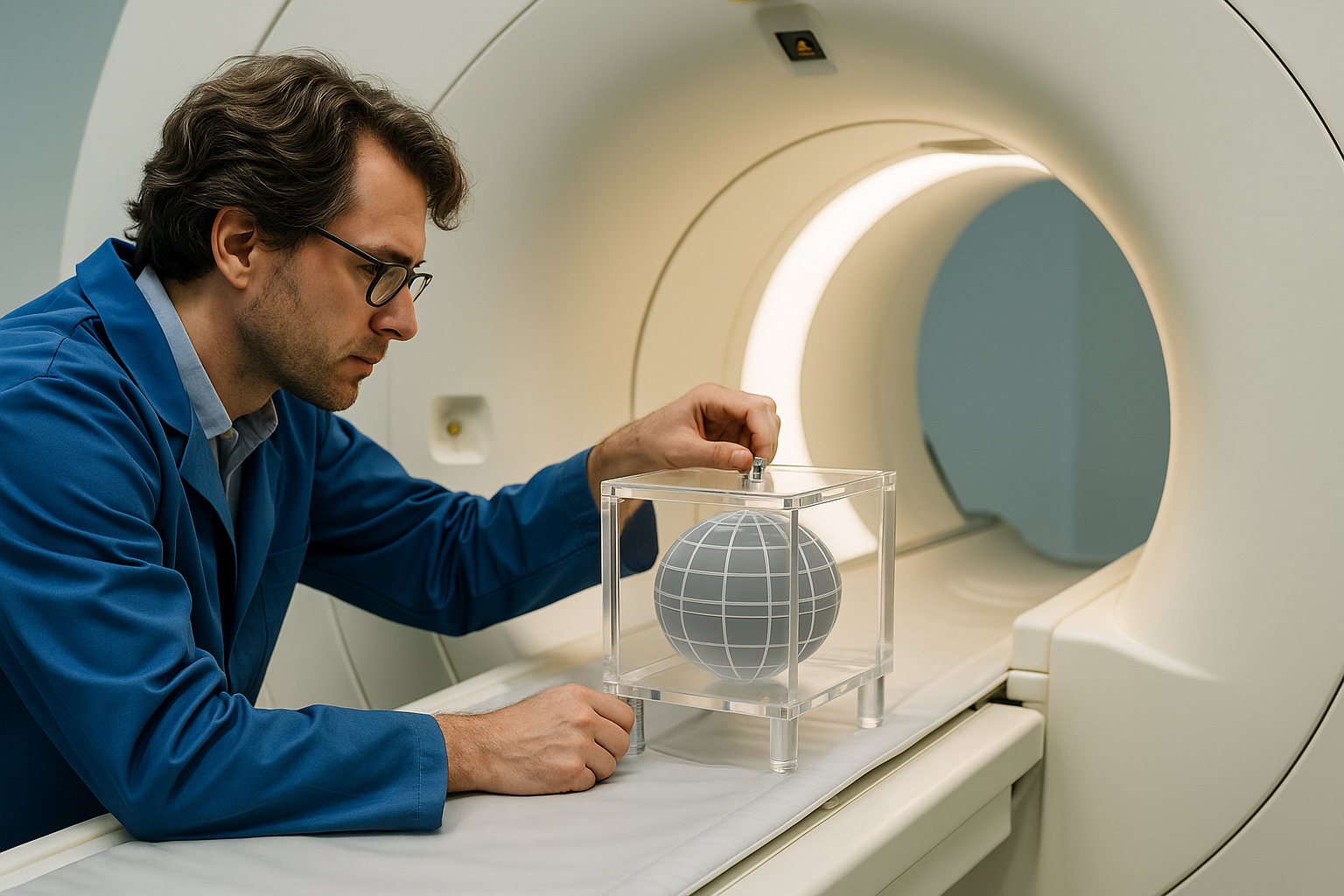
Physics-Driven Quality Assurance
Ensure consistent, high-fidelity scans with routine hardware checks, phantom testing, and protocol verification.
Learn More
Advanced Imaging & Post-Processing
Leverage cutting-edge sequences and reconstruction algorithms to capture richer tissue contrast and streamline data workflows.
Learn More
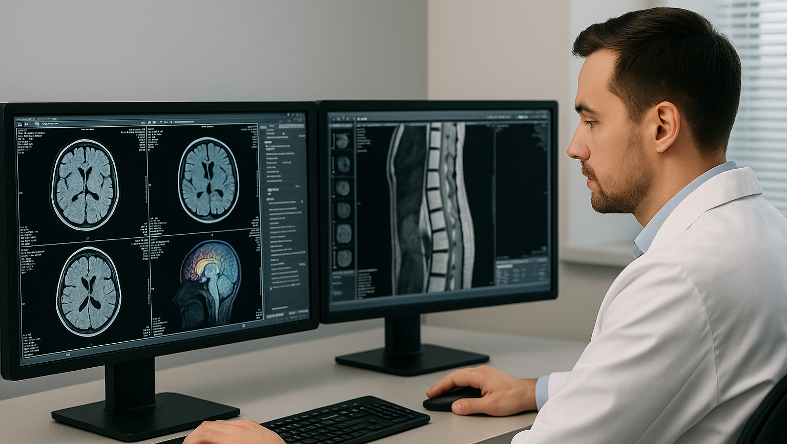
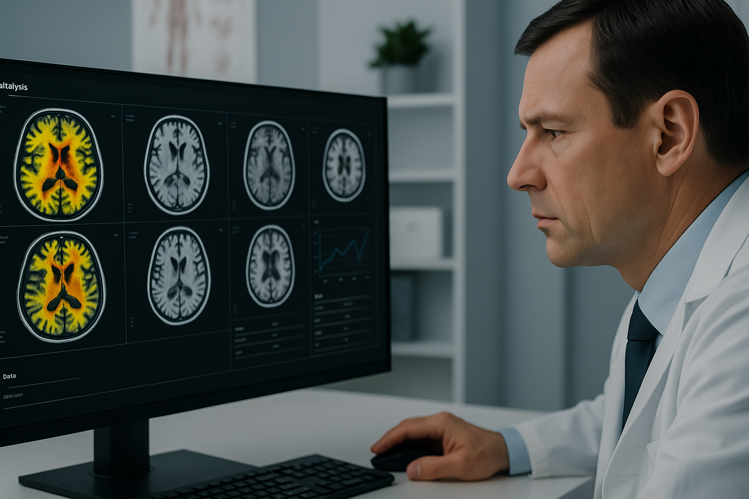
Clinical AI & Image Interpretation
Deploy validated AI modules for early disease screening and diagnostic support in neurology, oncology, and beyond.
Learn More