Automated MRI volumetry as a diagnostic tool for
Alzheimer’s disease
Automated MRI volumetry as a diagnostic tool for Alzheimer’s disease Structural neuroimaging with magnetic resonance imaging (MRI) (or computed tomography (CT)) plays a key role in the diagnostic work-up of dementia. It allows to rule out structural lesions of the brain that might cause cognitive problems. In addition, structural neuroimaging may contribute to the early and differential diagnosis of the neurodegenerative disease underlying the dementia syndrome. Applying automated measures of brain volumes on individual dementia patients requires a low measurement error and high reliability. Hence, the measurement error of the brain volumetric measures should be minimal, in order to draw meaningful conclusions in individual patients.
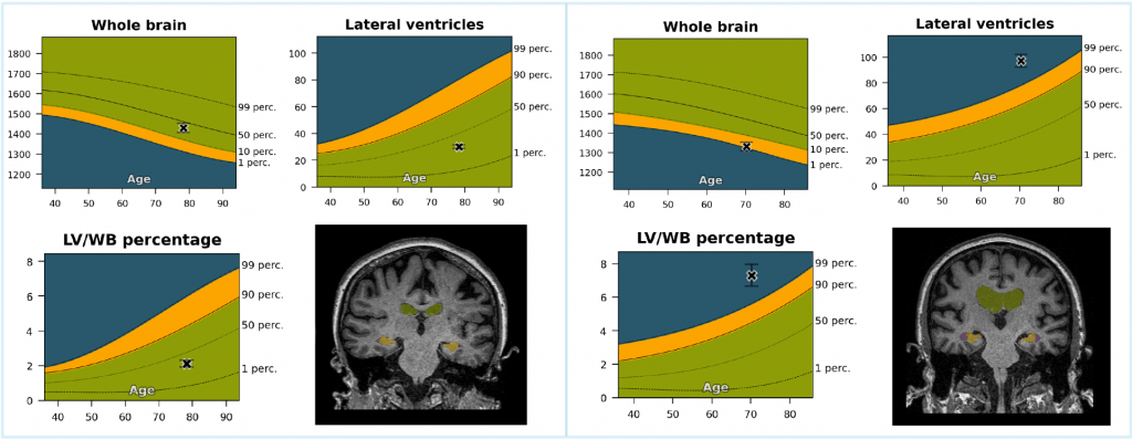
Whole-brain volume and ventricular enlargement
Whole-brain volume and ventricular enlargement are considered sensitive biomarkers of dementia due to their early and consistent involvement in neurodegeneration, supporting their utility for the timely diagnosis of AD. The relation between the whole brain and the ventricles can also be indicative of diseases such as normal pressure hydrocephalus (NPH). A high correlation is reported between the whole brain and ventricular volume and changes in cognitive performance in AD and MCI, and an even more robust correlation with their ratio.7 By using the ratio of measures, potential errors introduced by normalization methods are omitted, producing more statistically significant results for discriminating pathological subjects from normal controls.
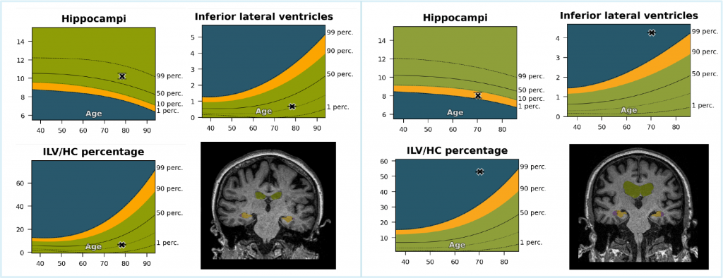
Hippocampal volume
Hippocampal volume loss is a supportive feature for the diagnosis of Alzheimer’s disease. However, visually disentangling age-related changes from pathological variations is challenging. Accordingly, the use of automated quantification software can help introduce hippocampal volume loss in the clinic. Neurodegeneration of the hippocampus (HC) typically co-occurs with the enlargement of the inferior lateral ventricle (ILV). This enlargement differentiates individuals with congenitally small hippocampi from those due to neurodegeneration and can act as an indicator for volume loss. The combination of hippocampal and inferior lateral ventricle volumes indicated the highest correlation with visual rating scales. Whereby the ILV/HC ratio has shown to have a small measurement error, which is crucial for detecting abnormalities during the radiological interpretation of MCI and AD.
Toward a timely and confident diagnosis
The use of automated quantification software can improve diagnostic sensitivity and accuracy while reducing the inter-and intra-rater variability for the detection of brain volume loss in clinical practice. Adding a volumetric quantitative analysis to standard brain MRIs, which are regularly used in diagnosing dementia patients, can therefore be a low-cost improvement toward a more timely and accurate diagnosis of Alzheimer’s disease.