Perfusion Imaging
Perfusion is a fundamental biological function that refers to the delivery of oxygen and nutrients to tissue by means of blood flow. Perfusion MRI is sensitive to microvasculature and has been applied in a wide variety of clinical applications, including the classification of tumors, identification of stroke regions, and characterization of other diseases. Perfusion MRI techniques are classified with or without using an exogenous contrast agent. Bolus methods, with injections of a contrast agent, provide better sensitivity with higher spatial resolution, and are therefore more widely used in clinical applications. However, arterial spin-labeling methods provide a unique opportunity to measure cerebral blood flow without requiring an exogenous contrast agent and have better accuracy for quantification. Importantly, MRI-based perfusion measurements are minimally invasive overall, and do not use any radiation and radioisotopes.
Magnetic resonance techniques have been powerful in visualizing tissue perfusion in the brain and other parts of body. Perfusion normally refers to the delivery of blood at the level of the capillaries, and measures in units of milliliters per 100 gram per minute. Perfusion is closely related to the delivery of oxygen and other nutrients to the tissue. Perfusion is, therefore, an essential parameter; and for this reason, much effort has been put into its measurement. It is important to distinguish between perfusion and bulk blood flow, which occurs along major arteries and veins.

Two main perfusion MRI approaches have been developed: those with and without the use of an exogenous contrast agent. The first group of techniques includes dynamic susceptibility contrast (DSC)-MRI and dynamic contrast enhanced (DCE)-MRI; while the second group relates to arterial spin-labeling (ASL). DSC-MRI is used only in the brain for the clinical evaluation of perfusion in cerebral ischemia and brain tumors. This technique involves the rapid intravenous injection of a magnetic resonance contrast agent and the serial measurement of signal loss during the passage of the bolus through the tissue, using T2 or T2* weighted images. DCE-MRI is another perfusion MRI method that relies on the injection of a contrast agent, but where T1-weighted magnetic resonance images are acquired dynamically before, during, and after bolus injection of a contrast agent. The data can be interpreted in terms of physiological tissue characteristics by applying the physical principles of tracer-kinetic modeling. This method has become standard in many applications. In contrast, ASL is a perfusion MRI method for quantitatively measuring cerebral perfusion, which is also referred to as cerebral blood flow (CBF), by taking advantage of using the magnetically labeled blood itself as an endogenous tracer. ASL has been extensively performed in the research arena, and sporadically applied in diseases.
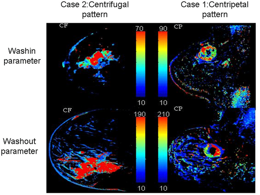
Contrast-based perfusion imaging methods require a high temporal resolution to capture the pass of the bolus, particularly when most of the contrast agent remains intravascular. These perfusion-imaging methods allow the estimation of several important hemodynamic parameters, which include blood flow, blood volume, and the mean transit time (MTT). So far, the major applications have been in the assessment and management of patients with acute stroke and tumors. measurement of contrast agent permeability, such as a transport constant related to the permeability surface area (Ktrans) and the fractional volume of the extravascular extracellular space (EES, ve), may be useful to evaluate diverse diseases.
What is a perfusion MRI scan? A brain perfusion scan is a type of brain test that shows the amount of blood taken up in certain areas of your brain. Other tests, such as the computed tomography (CT) perfusion or magnetic resonance imaging (MRI) perfusion, do not use radiotracers.
Dynamic Susceptibility Contrast (DSC)-MRI
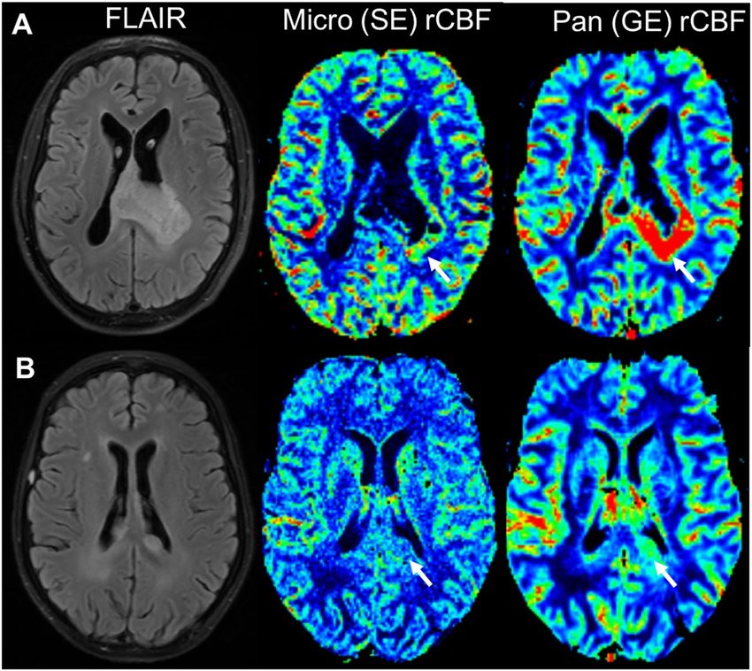
Dynamic susceptibility contrast-MRI is one of the exogenous contrast based methods, and relies on the intravenous injection of a paramagnetic contrast agent, such as those involving gadolinium (Gd) chelates, to generate a well-defined bolus. Most of the Gd chelates, e.g., Gd-diethylenetriaminepentacetate, are non-diffusible blood pool tracers. This technique utilizes very rapid imaging to capture the first pass of the contrast agent, and it is therefore also known as bolus tracking MRI. This technique is based on the susceptibility changes after injecting the contrast agent. The contrast agent is a paramagnetic material, which distorts the magnetic field, and reduces T2 around the vessel because of an increased susceptibility effect.
What is Perfusion used for? A brain perfusion scan is a type of brain test that shows the amount of blood taken up in certain areas of your brain. This can provide information on how your brain is functioning.
Dynamic Contrast-Enhanced (DCE)-MRI
Dynamic contrast-enhanced-MRI is the other exogenous contrast-based method. After the bolus of the contrast agent is injected, hemodynamic signals of DCE-MRI depend on the T1 relaxation time, and increase because of the T1 shortening effect associated with the paramagnetic contrast agent. DCE-MRI uses rapid and repeated T1-weighted images to measure the signal changes induced by the paramagnetic tracer in the tissue as a function of time. The relation between the DCE-MRI signal and T1 relaxation time is dependent on the details of the MR sequence used.
What does contrast enhanced MRI mean? Contrast enhancement is a ubiquitous term in radiology and can be used in three ways. Firstly, it may refer to any method of exaggerating the visible difference between adjacent structures on imaging by administering contrast media/agents. This includes differentiating between normal structures.
Arterial Spin Labeling (ASL)
Arterial spin labeling gives absolute values of perfusion of tissue by blood. This technique utilizes arterial water as an endogenous diffusible tracer, which is usually achieved by magnetically labeling the incoming blood. Therefore, ASL is completely noninvasive, using no injected contrast agent or ionizing radiation and is repeatable for studying normal or abnormal physiology and its variation with time.
What is ASL MRI? Arterial spin labeling (ASL) is a non-ionizing and completely non-invasive MRI technique for measuring tissue perfusion (blood flow), which uses magnetically labeled arterial blood water protons as an endogenous tracer.
