Diffusion Tensor Imaging (DTI) is an magnetic resonance imaging‑based neuroimaging technique that makes it possible to estimate the location, orientation, and anisotropy of the white matter tracts of the brain. (DTI) is an MRI technique that measures water molecule diffusion and its directionality. Because water diffusion in the brain is mainly restricted by fiber tracts, DTI has been suggested as an indirect marker for white-matter integrity. DTI might help identify and characterize epileptogenic lesions such as hippocampal sclerosis or FCD. DTI might also prove useful in the detection of lesions undetected by conventional MRI. With diffusion tensor imaging (DTI) it is possible to have in vivo localization of neuronal fiber tracts. DTI uses the anisotrophy inherent within white matter axons of the brain. Alignment of fiber tracts determines the water diffusion anisotrophy or directionality. Diffusion of water is preferential along the longitudinal axis of the axon; at the same time, the diffusion of water along the perpendicular axis is restricted. In DTI, each voxel has one or more pairs of parameters—a rate of diffusion and a preferred direction of diffusion—described in terms of three-dimensional space.
MR DTI allows identification and characterization of white matter tracts according to the direction and degree of their anisotropic water diffusion. Quantifying the degree of anisotropy in terms of metrics such as the fractional anisotropy offers insight into white matter development and degradation. There are fractional anisotropy changes in the white matter of brain neoplasms that might indicate cellular infiltration beyond the area of the tumor enhancement.
The DTI technique involves the delivery of external magnetic pulses to impose a random phase shift for water molecules that diffuse. This leads to a loss of signal from diffusing molecules, which subsequently creates darker volumetric pixels or voxels. For example, white matter fibers that are running parallel to the direction of the magnetic field gradient will produce a dark diffusion-weighted image for that particular direction. The diffusion tensor is then measured by comparing the signal loss with the original signal.
Two main parameters that are calculated from the tensor to define the nerve cell orientation are fractional anisotropy (FA) and mean diffusivity (MD). FA defines the degree of diffusion directionality, and MD provides the information on the average diffusivity of water.
Representation Of DT Images
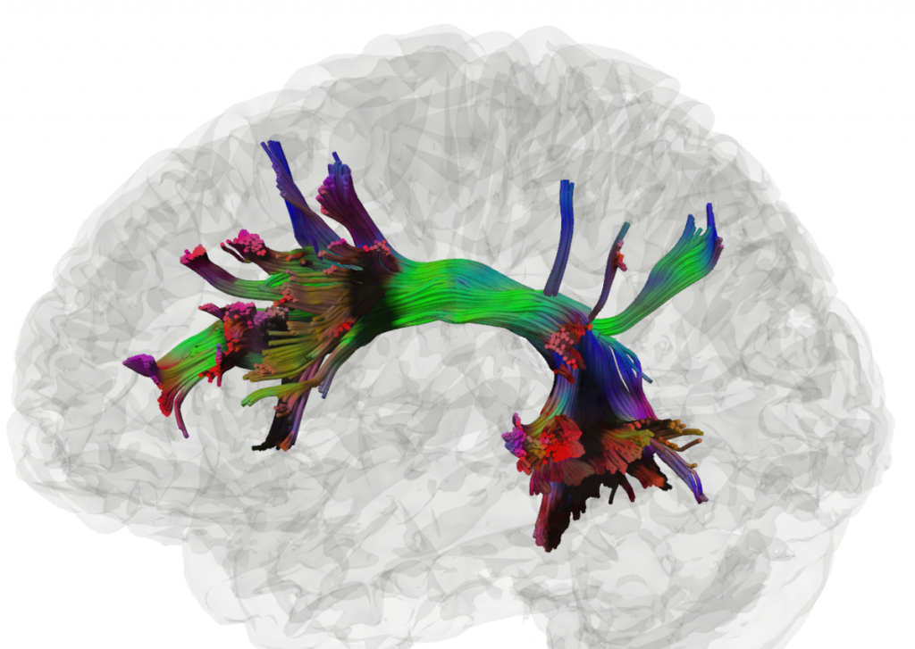
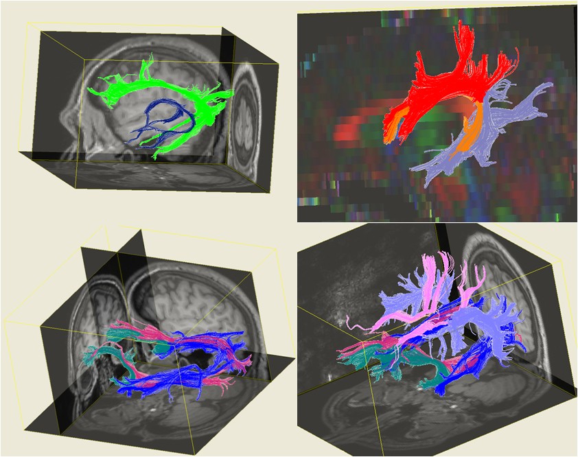
The information obtained from the tensor is either condensed into one number (scalar) or 4 numbers. Condensation into 4 numbers is performed to display the image with an R (red), G (green), B (blue) color and a brightness value.
Applications Neurophysiology
DTI is a very powerful technique for investigating important aspects of basic neurophysiology as well as pathophysiological consequences associated with disorders of the central nervous system. One major advantage is that information on the orientation of nerve fiber tracts can be easily obtained through this technique.
Brain Anatomy
In addition, the spatial distribution of complex brain network can also be investigated by measuring the microscopic length scale of water diffusion. As DTI provides additional information regarding the brain anatomy over conventional MRI, it is regarded as a more sensitive tool to investigate normal brain development as well as congenital brain development disorders.
Neuropathology
Since diffusion anisotropy is significantly related to the axon myelination status, measurement of this parameter using DTI can also provide substantial insight into neuropathological conditions related to demyelination, such multiple sclerosis.
Limitations
DTI provides a thorough anatomical overview of the white matter in terms of diffusion anisotropy and fiber orientation. However, some limitations do exist related to the interpretation of the data obtained. For instance, the three-dimensional diffusion model of DTI can characterize only one fiber in a volumetric pixel; thus, a visual representation of data obtained from a region of crossing fibers can be confusing. In other words, DTI can generate false positive or negative data in the case of brain regions that contain multiple fiber bundles oriented in different directions. Another limitation is that, since DTI is based on the diffusion of water molecules, it is not possible to differentiate axon directionalities through this technique.