Quantitative Volumetric Analysis for Brain MRI
Getting definitive answers is always important. Now more than ever it is crucial!
KIO Medical’s BioQUANT technologies is leading the way by utilizing quantitative volumetric MRI values to aid both physicians and their patients with quick, clear and objective feedback, and reproducible results for human brain atrophy assessment.
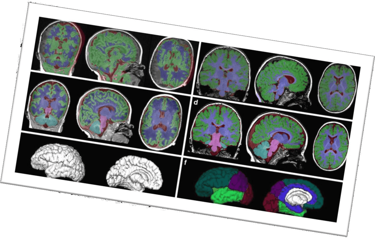
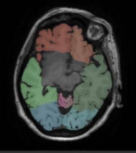
Automatic segmentation for different areas of the brain
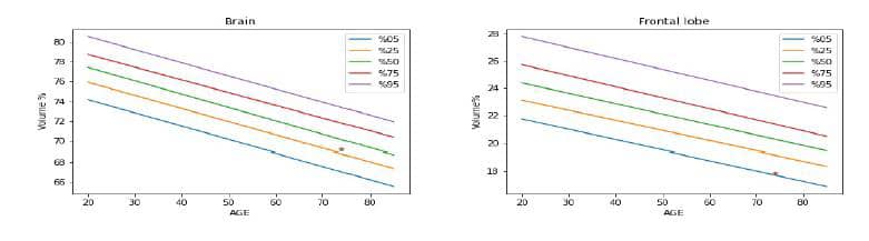
Leveraging a 3D T1-weighted scan, volumes of total brain, brain lobes (frontal, temporal, parietal and occipital), cerebellum and hippocampus are computed using state-of-the-art automatic segmentation techniques. These volumes can be compared to the reference values based on a healthy population study, or to a follow-up exam as described later.
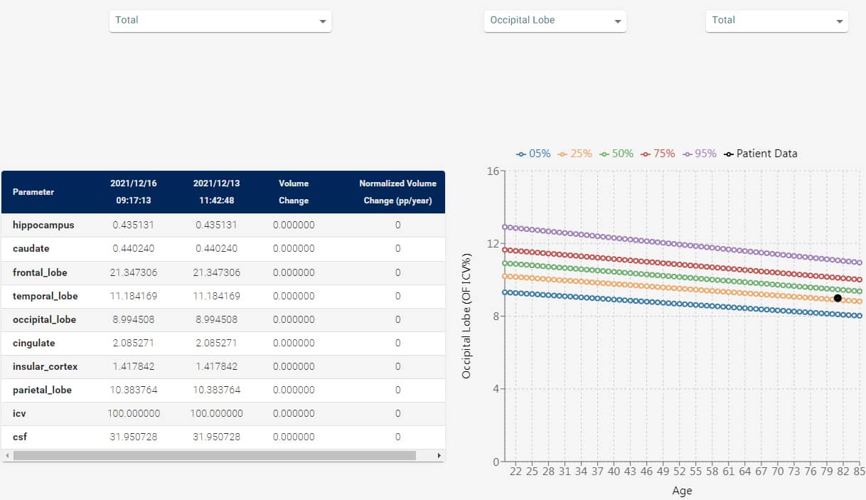
Follow up
If the patient has been scanned twice, it is possible to compare the follow-up scan with the baseline reference centile curves, allowing the user to compare the volume alterations between both scans based on the volume changes normalized to the reference curve models.
Workflow integration
Automatic segmentation of brain structures and atrophy assessment are now available without any excessive user interaction. It is therefore possible to automatically start the algorithms as soon as suitable scans are available at our cloud-based platform.
Disease
- Alzheimer’s Disease
- Focal Epilepsy
- Migraine
- Amyotrophic Lateral Sclerosis (ALS)
- Parkinson’s Disease
- Multiple Sclerosis
- Temporal Lobe Epilepsy (TLE)
- Mild Cognitive Impairment (MCI)
- Frontotemporal Dementia